Independent living and mobility is becoming a key condition for quality of life and social participation. Even though the musculoskeletal system is capable of fascinating complex actions, analogous to other systems it can break down due to dysfunction, wear or trauma. In my academic career, I have contributed by focusing on minimally invasive diagnosis and intervention and strived for the design of medical devices that form a ‘perfect’ synergy with the capabilities of the clinician. My approach has been ‘clinically driven’ and combining of (experimental) research, conceptual design and management of multidisciplinary teams. This has resulted in various public and public-private awarded (inter)national grants, over peer reviewed papers, several patents, and contributions to various start-ups. I find it very important that fruitful ideas in the end can actually be used by the clinicians to diagnose of treat patients in their best effort. This means that apart from prototyping and testing, attention needs to be paid to the business case and Medical Device Regulation (MDR). In my role of chair of Biomedical Device Design & Production, I aim to further develop my approach towards personalized & circular device design, shortening the development and validation phases and facilitating production of complex devices. Also, exploring the offering of medical devices via open source platforms while still complying to the MDR is performed.
The sharing of my knowledge I value highly, which is expressed in my active involvement in education of engineering and design courses and supervision of students. Also, I contribute(d) to the development of various bachelor and master programmes at University Twente, Delft University of Technology, Zuyd University of Applied Science and Maastricht University.
I am member of the Netherlands Academy of Engineering (NAE). More information: www.nae.nl
Expertise
Medicine and Dentistry
- Surgeon
- Computer Assisted Tomography
- Sacroiliac Joint Fusion
- Knee
- Osteotomy
- Surgical Planning
- Ankle Joint
- Range of Motion
Organisations
Ancillary activities
- RAAK SIAReview grant proposals
- DUTCHReview grant proposals
Publications
2026
2025
Research profiles
Affiliated study programs
Courses academic year 2026/2027
Courses in the current academic year are added at the moment they are finalised in the Osiris system. Therefore it is possible that the list is not yet complete for the whole academic year.
Courses academic year 2025/2026
Courses academic year 2024/2025
Current projects
Patient specific sacroiliac joint fusion to improve quality of life for woman with severe pelvic instability
PSI-FUSE
Severe pelvic instability causes severe pain and immobility. Stabilizing implants in the sacroiliac joint aim to reduce pain and restore function, but malpositioned implants pose risks of complications such as loosening and nerve damage. This study aims to determine the optimal patient-specific implant position using a finite element method. Subsequently, this optimal implant position can be achieved with a patient-specific surgical guide, of which a prototype has been developed. This will be validated by means of a cadaver study. Implementing this approach may reduce risks of complications and improve the quality of life for women with disabling pelvic instability.
Towards advanced 3D leg malalignment correction utilizing dynamic movement analysis
Mal-alignment of the lower limb can cause excessive stress on knee structures during movement. Traditionally, an osteotomy is performed to neutralize unbalanced forces, but is planned using alignment angles on a static radiograph taking no dynamic force aspects into account that occur during activities of daily living (ADL). Therefore, we aim to expand the target of a corrective osteotomy from a statically aligned limb to normalized internal loads within the knee occurring during various dynamic activities. Eventually, this approach will allow for movement-induced personalized alignment corrections, thereby reducing knee cartilage wear, anterior cruciate ligament re-rupture rate and failed return-to-sport.
Experimental Tissue Characterization: Cutting edge tool for science and education
Understanding human tissue properties plays a meaningful role in research and education of medical as well as engineering disciplines. Up till now, the teaching approach had been quite limited to demonstrating anatomies or calculating biomechanical properties, respectively. In both education models, room for enhanced and profound learning experiences is available. Research wise, data on human tissue properties are scarce, and if present, reported by limited statistical parameters, but certainly not location or functionality dependent parameters. The ambition is to fill the research and education gaps by developing a novel education module that uses hand-on measurements on human tissues toward enhanced learning experience while collecting high quality data for open access database. We envision to start with the development of one axial force measurement setup dedicated to tendons/ligaments, along with complementary standardized measurement protocols. The module allows UT/VU students to experience the look or feel of human tissue, perform experiments, process data, perform (bio)statistics, and report according to research standards. As a pilot, we intend to incorporate the novel module in a new course-Medical Device Prototyping, which will be an elective course for UT/VU students. Once this education module is established, we explore integration of it in other courses such as the ones that academic teaching and research skills are among the learning goals. The module itself will be scaled up with additional experimental setups (e.g. biaxial and shear forces) and tissue types. The setups are suitable for joint master assignments to compare properties of fresh frozen and embalmed (Fix for life method) cadaver tissues. The continued generation of reliable human tissue data will be valuable for research areas such as virtual twin models, life-like training and education phantoms, surgical instrument and implant development.
H3D- VISIOnAiR
Head-worn 3D-Visualisation of the Invisible for Surgical Intra-Operative Augmented Reality
During surgery surgeons must identify vital anatomical structures (nerves and lymph nodes) to prevent unnecessary healthy tissue damage. Correct intraoperative identification by normal eyesight remains enormously challenging due to natural variation. Therefore, surgeons require high-tech intra-operative imaging for reliable high-resolution visual discrimination of critical anatomical structures. H3D-VISIONAIR offers a breakthrough and disruptive head-worn augmented reality (AR) system for surgeons to see beyond normal eyesight. It consists of two multi-spectral cameras (combining visual range with NIR visualization), a belt computer with real-time data processing, and a high-end stereoscopic head mounted display with wireless connection to the operating room infrastructure. The spectral signature of specific annotated tissues form input to develop machine-learning based models to allow automatic segmentation. The models are used to generate the AR-overlays on top of the normal surgical field of view.
One-stop breath analyser
Decreased deployment of medical resources by applying the one-stop breath analyser in vulnerable patients with chronic diseases
Early diagnosis of chronic diseases in children and elderly is crucial to improve treatment and prognosis. Non-invasive analysis of exhaled breath has shown potential for improved diagnosis and management and our TRL4 lab prototype breath analyser with carbon nanotube sensors is further developed for this purpose into a TRL6 prototype.
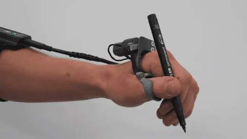
Thumbs up!
Development of the T-Grip assistive device
The State of the Cartilage (START)
Marco van Basten’s case is puzzling: why does a healthy, active teenager end up with a lasting disability from ankle osteoarthritis just a few years after a simple ankle sprain? To prevent this from happening again, we are developing a new tool that can measure whether there is damage to the ankle and how severe it is immediately after the injury. In a large-scale study, we want to find out if this damage gets worse, leading to osteoarthritis. With this tool and what we learn, we hope to be able to treat osteoarthritis before it reaches the point of no return.
Dynamic smart implant to permanently correct scoliosis
DYNACURE
Scoliosis is a deformation of the spine that affects 2-5% of teenagers. When severe, the spine is fixed with rods, which limits trunk motion. The aim is to correct the scoliosis without limits. As basis, we use our pre-existing implant that already allows for growth and motion. This implant is made smart by integrating a mechanism that adjust its loading to the invidual patient situation. The level of adjustment is determined with a computer model that predicts optimal loading for correction over time. The novel implant ultimately permantly corrects scoliosis. Together with UMCU, TU Eindhoven, Cresco Spine and InSpine we will work on this important topic for the coming years
Finished projects
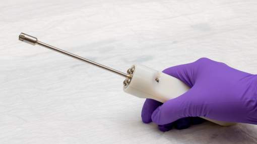
Double Spring Reduction system
Non-fusion scoliosis correction implant (TTT Voucher)
In this project, we aim to develop the current prototype (TRL5) further to make it ready for a pilot study in humans. The redesign is targeted to the fixation system as the screws that were used are no longer available on the market. With the redesign, Medical Device Regulation documents are generated as well as the IMDD to acquire METC permission. In the future, after successful clinical testing, a start-up company will bring the system to the market.
Waterjetdissection
TTT Voucher to explore valorization
The potential of waterjet dissection for targeted orthopedic surgery has been proven by a consortium of TU Delft, Amsterdam University Medical Centre, Maastricht University and University of Twente (UT) via a TRL 5 demonstrator. Lead by Maastricht Instruments and UT we explore its application and associated design changes in a business case to allow optimal execution of arthroplasty and revision surgery
Address
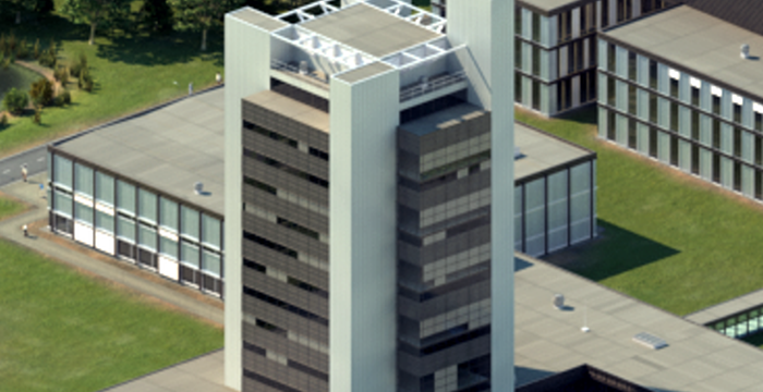
University of Twente
Horst Complex (building no. 20), room W113
De Horst 2
7522 LW Enschede
Netherlands
University of Twente
Horst Complex W113
P.O. Box 217
7500 AE Enschede
Netherlands
 Scan the QR code or